Monday 26 February 2024

Diabetic Retinopathy Detection Using Image Processing Matlab Project With Source Code Final Year Project

ABSTRACT

          The processing of images by performing some operations in order to get enhanced images is called as image processing. It is widely used to diagnose the eye diseases in an easy and efficient manner. Several techniques has been developed for the early detection of DR on the basis of  features  such as blood. It includes the image enhancement  processes  like  histogram  equalization  and adaptive histogram equalization for the detection of DR. The persistent  damage  caused  to  the  retina  is  termed  as  the retinopathy.  The  condition  of  diabetic  retinopathy  (DR) happens  with  those  who  have  diabetes  that  results  in progressive damage to the retina.  Due to high blood glucose levels it  leads to the  damage of small blood vessels in  the retina and this may result into swelling of the retina. ie., DR is a diabetes related eye disease which occurs when the blood vessels in the retina become swelled and leaks fluid which ultimately leads to vision loss. The DR is regarded as a serious sight threatening condition. The  main  objective of  this method  is to  detect DR (Diabetic  Retinopathy)  eye  disease  using  Image  Processing techniques. The tool used  in this method is MATLAB and it is widely used in image processing. This project proposes a method for Extraction of Blood Vessels from the medical image of human  eye-retinal  fundus  image  that  can  be  used  in ophthalmology  for  detecting  DR.  This  method  utilizes  an approach  of  Adaptive  Histogram  Equalization  using  CLAHE (Contrast  Limited Adaptive  Histogram Equalization)  algorithm with Convolutional Neural Networks algorithm implementation. The result shows that affected DR is detected in fundus image and the DR  is  not  detected  in  the  healthy  fundus  image  and upto 98%  of Accuracy can be achieved in the detection of DR Project. This project is developed in matlab.

PROJECT OUTPUT


PROJECT DEMO VIDEO

Contact:
Prof. Roshan P. Helonde
Mobile: +91-7276355704
WhatsApp: +917276355704
Email: roshanphelonde@rediffmail.com

Image Steganography Using DCT Algorithm For Hiding Data In Image Matlab Project With Source Code Final Year Project

ABSTRACT

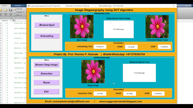
            Steganography is the science and art of secret communication between two sides that attempt to hide the content of the message. It is the science of embedding information into the cover image without causing a loss in the cover image after embedding. Steganography is the art and technology of writing hidden messages in such a manner that no person, apart from the sender and supposed recipient, suspects the lifestyles of the message. It is gaining huge attention these days as it does now not attract attention to its information's existence. In this project the secret message is encrypted first then DCT technique is applied. Moreover, Discrete Cosine Transform (DCT) is used to transform the image into the frequency domain.  DCT algorithm is implemented in frequency domain in which the stego-image is transformed from spatial domain to the frequency domain and the payload bits are inserted into the frequency components of the cover image.

PROJECT OUTPUT


PROJECT DEMO VIDEO

Contact:
Prof. Roshan P. Helonde
Mobile: +917276355704
WhatsApp: +917276355704
Email: roshanphelonde@rediffmail.com

Image Steganography Using Spread Spectrum Technique Matlab Project With Source Code Final Year Project

ABSTRACT

            Steganography is the science and art of secret communication between two sides that attempt to hide the content of the message. It is the science of embedding information into the cover image without causing a loss in the cover image after embedding. Steganography is the art and technology of writing hidden messages in such a manner that no person, apart from the sender and supposed recipient, suspects the lifestyles of the message. It is gaining huge attention these days as it does now not attract attention to its information's existence. Spread spectrum is the technique of modulating a narrow band signal with a wideband signal so that the energy of the resultant signal is spread evenly across a wider spectrum and therefore hard to detect. Spread Spectrum Image Steganography (SSIS) typically modulates the binary message signal into a noise signal and the result is added to an image. This resultant signal is similar to noise. By carefully adjusting the power of the embedded signal it is possible to achieve both imperceptibility of the hidden message and reasonable recovery of the signal. The stego-images produced by SSIS will be indistinguishable from images corrupted by noise. An error correcting code is incorporated into the embedding so that the errors in signal recovery can be corrected.

PROJECT OUTPUT


PROJECT DEMO VIDEO

Contact:
Prof. Roshan P. Helonde
Mobile: +917276355704
WhatsApp: +917276355704
Email: roshanphelonde@rediffmail.com

Leukemia Blood Cancer Detection Using CNN Convolutional Neural Network Matlab Project Source Code | Final Year Project

  ABSTRACT

             Leukemia Blood cancer is the most prevalent and it is very much dangerous among all type of cancers. Early detection of blood cancer has the potential to reduce mortality and morbidity. There are many diagnostic technologies and tests to diagnose blood cancer. However many of these tests are extremely complex and subjective and depend heavily on the experience of the technician. To obviate these problems, image processing techniques is use in this study as promising modalities for detection of Leukemia blood cancer. The accuracy rate of the diagnosis of blood cancer by using image processing will be yield a slightly higher rate of accuracy then other traditional methods and will reduce the effort and time. We first discuss the preliminary of cell biology required to proceed to implement our proposed method. This project presents a new automated approach for blood Cancer detection and analysis from a given photograph of patient’s cancer affected blood sample. The proposed method is using image improvement, image segmentation for segmenting the different cells of blood, edge detection for detecting the boundary, size, and shape of the cells and finally Clustering for final decision of blood cancer based on the number of different cells and classification done using convolutional neural network. This project is developed in matlab.

PROJECT OUTPUT


PROJECT DEMO VIDEO

Contact:
Prof. Roshan P. Helonde
Mobile: +917276355704
WhatsApp: +917276355704
Email: roshanphelonde@rediffmail.com

Currency Recognition Using Image Processing Matlab Project Source Code Final Year Project

ABSTRACT

             The Reserve Bank is the one which issue bank notes in India. Reserve Bank, changes the design of bank notes from time to time. The appearance of the currency is part of this development and it is affected directly, where there is exploited in incorrect form by copying the currency in a manner similar to the reality. Therefore, it became necessary to implement a proposal for being a suitable as solution not inconsistent with the different cultures, time and place. This clear through add the watermarks inside currency, which is difficult to be copied. At the same time, this watermarks may be visible to the naked eye so can easily inferred or it is invisible. However the high resolution imaging devices can copy these additions. In this research, we have proposed a system to distinguish the currencies by the program that working a submission inferred to the watermark by feature extraction determined the type of currency. In addition to, it determined category of the currency. Benefit of it, is reducing as much as possible the spread of counterfeit currency and this system can be used by any user wants to make sure of the currency. This project is developed in matlab.

PROJECT OUTPUT


PROJECT DEMO VIDEO
Contact:
Prof. Roshan P. Helonde
Mobile: +917276355704
WhatsApp: +917276355704
Email: roshanphelonde@rediffmail.com

Lung Cancer Detection Using Image Processing Matlab Project With Source Code | Final Year Project Code

 ABSTRACT

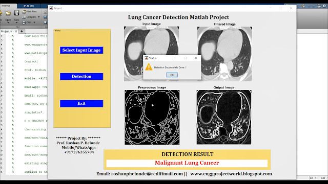
             Lung cancer prevalence is one of the highest of cancers. One of the first steps in lung cancer diagnosis is sampling of lung tissues or biopsy. These tissue samples are then microscopically analyzed. This procedure is taken once imaging tests indicate the presence of cancer cells in the chest. Lung cancer diagnosis using lung images. One of them is that doctor still relies on subjective visual observation. A medical specialist must do thorough observation and accurate analysis in detecting lung cancer in patients. Hence, there is need for a system that is capable for detecting lung cancer automatically from microscopic images of biopsy. This method will improve the efficiency for lung cancer detection. The aim of this project is to design a lung cancer detection system based on analysis of ct image of lung using digital image processing. A medical specialist must do thorough observation and accurate analysis in detecting lung cancer in patients. Hence, there is need for a system that is capable for detecting lung cancer automatically from images of lungs. This method will improve the efficiency for lung nodule detection. The aim of this project is to detect a lung cancer detection system based on analysis of lung images using digital image processing. Lung Cancer Detection done using image processing. This project is developed in matlab.

PROJECT OUTPUT


PROJECT DEMO VIDEO

Contact:
Prof. Roshan P. Helonde
Mobile: +91-7276355704
WhatsApp: +91-7276355704
Email: roshanphelonde@rediffmail.com

Saturday 24 February 2024

Image Watermarking Using DWT Discrete Wavelet Transform Algorithm Matlab Project With Source Code | Final Year Project Code

 ABSTRACT

          The use of Internet technology has led to the availability of different multimedia data in various formats. The unapproved customers misuse multimedia information by conveying them on various web objections to acquire cash deceptively without the first copyright holder’s intervention. Due to the rise in cases of COVID-19, lots of patient information are leaked without their knowledge, so an intelligent technique is required to protect the integrity of patient data by placing an invisible signal known as a watermark on the medical images. In this project image watermarking is proposed using discrete wavelet transform algorithm on both standard and medical images. The project addresses the use of digital rights management in medical field applications such as embedding the watermark in images. The various quality parameters are used to figure out the evaluation of the developed method. This project is developed in matlab.

PROJECT OUTPUT


PROJECT DEMO VIDEO

Contact:
Prof. Roshan P. Helonde
Mobile: +91-7276355704
WhatsApp: +917276355704
Email: roshanphelonde@rediffmail.com

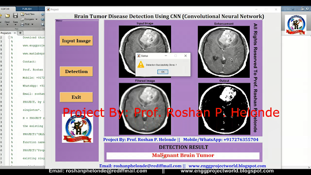
Brain Tumor Detection Using Convolutional Neural Network CNN Matlab Project With Source Code | Final Year Project

ABSTRACT

           Brain tumors are the most common issue in children. Approximately 3,410 children and adolescents under age 20 are diagnosed with primary brain tumors each year. Brain tumors, either malignant or benign, that originate in the cells of the brain. The conventional method of detection and classification of brain tumor is by human inspection with the use of medical resonant brain images. But it is impractical when large amounts of data is to be diagnosed and to be reproducible. And also the operator assisted classification leads to false predictions and may also lead to false diagnose. Medical Resonance images contain a noise caused by operator performance which can lead to serious inaccuracies classification. Brain tumor identification is really challenging task in early stages of life. But now it became advanced with various machine learning algorithms. Now a day’s issue of brain tumor automatic identification is of great interest. In Order to detect the brain tumor of a patient we consider the data of patients like MRI images of a patient’s brain. Here our problem is to identify whether tumor is present in patients brain or not. It is very important to detect the tumors at starting level for a healthy life of a patient. There are many literatures on detecting these kinds of brain tumors and improving the detection accuracies. In this work we used Brain Tumor Detection Using Convolutional Neural Network CNN. This project is developed in matlab. 

PROJECT OUTPUT


PROJECT DEMO VIDEO

Contact:
Prof. Roshan P. Helonde
Mobile: +91-7276355704
WhatsApp: +917276355704
Email: roshanphelonde@rediffmail.com

Acne Disease Detection Using CNN Convolutional Neural Network Python Project With Source Code | Final Year Project

ABSTRACT

          Acne is a chronic skin disease occurring from inflammation of pilosebaceous units which are hair follicles under skin and their surrounding sebaceous gland (fatty gland) clog up. Currently, dermatologist has to manually mark a location of acnes on the sheet, then count to quantify and measure treatment progress. This is an unreliable and inaccurate method. Moreover, this method requires dermatologist’s excessive effort. In this project, a novel automatic acne disease detection using Image processing technique is proposed. Acne causes significant physical and psychological problems for patients such as permanent scarring, depression and anxiety from poor self-image. When you have acnes, go to see dermatologist early is the safest way to heal and prevent future permanent scars. Acne can be caused by many factors such as overactive oil glands that produce too much oil, combine with skin cells to make pores in the skin, become plugged and p-acne bacteria cause acne disease. This project is developed in python using convolutional neural network.

PROJECT OUTPUT


PROJECT DEMO VIDEO
Contact:
Prof. Roshan P. Helonde
Mobile: +91-7276355704
WhatsApp: +917276355704
Email: roshanphelonde@rediffmail.com

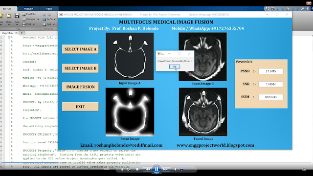
Multifocus Medical Image Fusion Using Matlab Project With Source Code | Final Year Project Code

ABSTRACT

             Fusion of image is a new discipline that involves the fusing of pictures collected by multiple sensors to generate an informative image which helps in medical diagnosis for making appropriate decisions. Image fusion is becoming increasingly important in computer vision activities because of the larger number of capture methods. The integration of different views like multi view, multi temporal view and information which is larger together termed as image fusion. In image fusion the information of multiple-sensors are converted into a unified image while maintaining the fidelity of critical characteristics. Healthcare image fusion procedures are used to improve picture quality by achieving the conspicuous characteristics in the fusion findings. As a result, they increase the practical usefulness of medical pictures for issue assessment and identification. Fusion of medical images are generally evaluated using the modalities like MRI-Magnetic Resonance Imaging and CT-Computed Tomography. Fusion of images is a compelling method for breaking down and utilizes enormous volumes of pictures near source in light of the fact that the fused picture incorporates different data information that can't be acquired from a solitary source picture. Accordingly, picture combination is utilized in a wider scope of utilization and some of them are clinical treatment, photography, remote detecting, military, security, and observation. This project is developed in matlab.

PROJECT OUTPUT


PROJECT DEMO VIDEO

Contact:
Prof. Roshan P. Helonde
Mobile: +91-7276355704
WhatsApp: +917276355704
Email: roshanphelonde@rediffmail.com

Medical Image Encryption and Decryption Matlab Project With Source Code | Medical Data Encryption and Decryption

 ABSTRACT

             In recent years mostly all the health centers and hospitals use the wireless networks and internet for biomedical information exchanging, the secure of this information in not verified and cannot be grantee in such environment, the personality of patient and for security concerns inside such institutions there is a need for encryption system that can easily encrypt the biomedical data and it can be shared with other centers via internet without and concerns about privacy. Our system based on advanced encryption standard with encryption and decryption taking to consideration the criticality of data that been encrypted. Medical image security is very important issue in new world technologies with the internet of things revolution everything is connected to the internet and need to protected and authenticated, our project can encrypt the medical images for popular people or the critical situation patient that can help to protect the patients privacy by merging many techniques.

PROJECT OUTPUT


PROJECT DEMO VIDEO
Contact:
Prof. Roshan P. Helonde
Mobile: +91-7276355704
WhatsApp: +917276355704
Email: roshanphelonde@rediffmail.com

Audio Steganography Hiding Secret Text In Audio Matlab Project With Source Code | Final Year Project

ABSTRACT

             Steganography is one of the best data hiding technique in the world which can be used to hide data without its presence felt. In today’s digital world most of us communicate via use of electronic media or internet. Most people among us remain unaware about the data loss or data theft which can happen on online transmission of data or message. Valuable information including personal data, messages transmitted through internet is vulnerable to hackers who may steal or decrypt our data or messages. This poject is about enhancing the data or message security with use of Audio Steganography using LSB algorithm to hide the message into multiple audio files. The message hidden by this application is less vulnerable to be stolen than other similar applications. This is due to following reasons: Firstly files are taken to hide high amount of message which enhance information hiding capacity. Secondly before being hidden, the message is broken into parts and shuffled randomly based on permutation generated at runtime so even if the Least Significant Bit gets encountered the message is still unarranged and meaningless which enhances its security. This application is capable to carry large amount of information with greater security. As audio steganography uses audio as a cover medium, similarly this application too uses an audio as a platform for hiding the message. User provides input message in the form of text and chooses the audio wave file to hide the message. This application provides a smart and interactive interface for message hiding and its retrieval. Message is shuffled in random sequence before being hidden. Random sequence which is generated based on certain factors is used to shuffle the message before hiding it. This further enhances the data security. 

PROJECT OUTPUT


PROJECT DEMO VIDEO

Contact:
Prof. Roshan P. Helonde
Mobile: +917276355704
WhatsApp: +917276355704
Email: roshanphelonde@rediffmail.com

Wednesday 21 February 2024

Maize Leaf Disease Detection Using Image Processing Matlab Project With Source Code | Final Year Project Code

 ABSTRACT

         Crop disease protection is important for global food security, while the recognition of crop diseases at early stage is the key part of disease protection. The traditional identification and detection of crop leaf diseases is carried out by agricultural technicians. The identification and diagnosis of crop leaf disease is of great significance to improve the quality of crop cultivation. Compared with the traditional manual diagnosis method, the automatic identification of crop leaf disease based on computer vision technology has high efficiency and no subjective judgment error. But the traditional image processing technology is affected by different illumination conditions, cross shading. The algorithm's robustness is affected. Because deep learning dose not need to set learning features manually, which greatly improves the recognition efficiency. This project is developed in matlab using image processing.

PROJECT OUTPUT

PROJECT DEMO VIDEO
Contact:
Prof. Roshan P. Helonde
Mobile: +91-7276355704
WhatsApp: +917276355704
Email: roshanphelonde@rediffmail.com

Glaucoma Detection Using Convolutional Neural Network Matlab Project With Source Code | Final Year Project Code

ABSTRACT

                Computational techniques have great impact in the field of Medicine and Biology. These techniques help the medical practitioners to diagnose any abnormality in advance and provide fruitful treatment. Retinal image analysis has been an ongoing area of research. Automated retinal image analysis aid the ophthalmologists in detecting abnormalities in the retinal structures namely optic disc, blood vessels, thus diagnosing sight threatening retinal diseases such as Glaucoma and Retinopathy. Glaucoma is the major cause of blindness in working population. Glaucoma is characterized by increased intra-ocular pressure inside the eye leading to changes in the optic disc and optic nerve. It does not reveal its symptoms until later stage. Hence, regular screening of the patients is required to identify the disease, thus demanding high labor, time and expertise. Thus, computational techniques are sought for their analysis. In this project, identification of Glaucoma is carried out through computational techniques namely image processing. As the changes in the profile of optic disc act as a biomarker for the onset of the disease, optic disc is segmented through image processing techniques. Optic disc is the brightest part portrayed as oval structure in the retinal fundus image. It encompasses optic cup, which is the brightest central part, optic rim, the surrounding pale part and the blood vessels. All these structures are segmented and their properties are elicited. Then, properties of the disc, cup and blood vessels within optic disc are mined to design a learning model for prediction of Glaucoma. This project is developed in matlab.

PROJECT OUTPUT


PROJECT DEMO VIDEO

Contact:
Prof. Roshan P. Helonde
Mobile: +91-7276355704
WhatsApp: +917276355704
Email: roshanphelonde@rediffmail.com

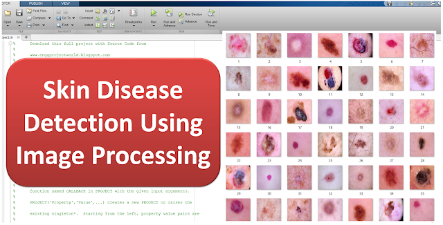
Skin Disease Detection Using Image Processing Matlab Project With Source Code | Final Year Projects

ABSTRACT

         Skin disease also known as melanoma it is one of the deadliest form of disease if not recognized in time. Since the pigmented areas/moles of the skin can be nicely observed by simple, non-invasive visual inspection the clinical protocols of its recognition also consider several visual features. Melanoma is the deadliest form of skin cancer, which is considered one of the most common human malignancies in the world. Early detection of this disease can affect the result of the illness and improve the chance of surviving. The tremendous improvement of deep learning algorithms in image recognition tasks promises a great success for medical image analysis, in particular, melanoma classification for skin disease diagnosis. Activation functions play an important role in the performance of convolutional neural networks for image recognition problems as well as medical image classification. Melanin is the pigment that discerns the color of human skin. The special cells produce melanin in the skin. If these cells are damaged or unhealthy, skin discoloration is visible. Skin pigment discoloration on cheeks is a hazardous fact as a symptom of human skin disease with a possibility of losing natural beauty. The extracted information of the skin discoloration can work as a guide to diagnosis the disease. In this project, different imaging techniques like preprocessing method and classification are used to analyze and extract the information of skins discoloration disease from skin images. This project is developed in matlab.

PROJECT OUTPUT


PROJECT DEMO VIDEO

Contact:
Prof. Roshan P. Helonde
Mobile: +91-7276355704
WhatsApp: +917276355704
Email: roshanphelonde@rediffmail.com

Shadow Detection and Removal Using Image Processing Matlab Project With Source Code | Final Year Project

 ABSTRACT

           Shadow Detection and removal from images is a challenging task in visual surveillance and computer vision applications. The appearance of shadows creates severe problems. Shadows cause problems in PC vision and image processing, such as detection of edge, video surveillance, stereo registration, object recognition, and image segmentation .To identify and remove the shadows from the image gives practical significance in image processing. Shadow forms when direct light cannot reach properly. This project proposes a simple method to detect and remove shadows from a single RGB image. A shadow detection method is selected on the basis of the mean value of RGB image in A and B planes of LAB equivalent of the image and shadow removal method is based on the identification of the amount of light impinging on a surface. The lightness of shadowed regions in an image is increased and then the color of that part of the surface is corrected so that it matches the lit part of the surface.

PROJECT OUTPUT


PROJECT DEMO VIDEO

Contact:
Prof. Roshan P. Helonde
Mobile: +91-7276355704
WhatsApp: +917276355704
Email: roshanphelonde@rediffmail.com

Skin Disease Detection Using CNN Python Project With Source Code | Skin Disease Classification Using CNN

 ABSTRACT

         Skin disease also known as melanoma it is one of the deadliest form of disease if not recognized in time. Since the pigmented areas/moles of the skin can be nicely observed by simple, non-invasive visual inspection the clinical protocols of its recognition also consider several visual features. Melanoma is the deadliest form of skin cancer, which is considered one of the most common human malignancies in the world. Early detection of this disease can affect the result of the illness and improve the chance of surviving. The tremendous improvement of deep learning algorithms in image recognition tasks promises a great success for medical image analysis, in particular, melanoma classification for skin disease diagnosis. Activation functions play an important role in the performance of convolutional neural networks for image recognition problems as well as medical image classification. Melanin is the pigment that discerns the color of human skin. The special cells produce melanin in the skin. If these cells are damaged or unhealthy, skin discoloration is visible. Skin pigment discoloration on cheeks is a hazardous fact as a symptom of human skin disease with a possibility of losing natural beauty. The extracted information of the skin discoloration can work as a guide to diagnosis the disease. In this project, different imaging techniques like preprocessing method and classification are used to analyze and extract the information of skins discoloration disease from skin images. This project is developed in python.

PROJECT OUTPUT


PROJECT DEMO VIDEO

Contact:
Prof. Roshan P. Helonde
Mobile: +917276355704
WhatsApp: +917276355704
Email: roshanphelonde@rediffmail.com

Diabetic Retinopathy Detection Using CNN Convolutional Neural Network Matlab Project With Source Code Final Year Project

 ABSTRACT

           Diabetic Retinopathy (DR) is a chronic health disease which requires early detection and treatment. It is important to identify DR using an intelligent system for faster prediction since manual examination and detection of the disease are unreliable and highly prone to error. Therefore, various researchers and medical experts have adopted and approached for advanced feature extraction and image classification, for early DR detection. Diabetic Retinopathy is a consequence of diabetes that affects the eyes. Damaged blood vessels in the retina, a light-sensitive tissue, are the primary cause of DR. If the patient has a long-term case  of diabetes and  the blood sugar  level is  not regulated consistently, the odds of this  issue developing in the eye increase.  Diabetic  Retinopathy is  one  of  the most  common causes  of  blindness  in  the Western  countries. Preventing Diabetic Retinopathy has  found to be quite beneficial when people with  diabetes are  monitored regularly. This  process is  shown to be essential if Diabetic Retinopathy is discovered in its early stages due to the availability of treatment. Diabetic Retinopathy, the main cause of blindness among working-age adults, affects millions of individuals. Diabetic  Retinopathy  is  a  medical  disorder  in  which  diabetes  mellitus  causes  damage  to  the  retina.  Diabetic Retinopathy  is diagnosed  using  colored  fundus  images,  which  requires  trained clinicians  to recognize  the  presence  and importance  of  several tiny  characteristics,  making  it a  time-consuming  task.  We present  a convolutional neural network CNN based  technique to  detect diabetic retinopathy in fundus images in this project. This project is developed in matlab.

PROJECT OUTPUT


PROJECT DEMO VIDEO
Contact:
Prof. Roshan P. Helonde
Mobile: +917276355704
WhatsApp: +917276355704
Email: roshanphelonde@rediffmail.com

Medical Image Fusion Using Curvelet Transform Technique Matlab Project with Source Code Final Year Major Project

 ABSTRACT

               Image fusion is the process of merging two images of the same scene to form a single image with as much information as possible. Image fusion is important in many different image processing fields such as satellite imaging, remote sensing and medical imaging. The study in the field of image fusion has evolved to serve the advance in satellite imaging and then, it has been extended to the field of medical imaging. Several fusion algorithms have been proposed extending from the simple averaging to the curvelet transform. The wavelet fusion algorithm has succeeded in both satellite and medical image fusion applications. The basic limitation of the wavelet fusion algorithm is in the fusion of curved shapes. Thus, there is a requirement for another algorithm that can handle curved shapes. So, the application of the curvelet transform for curved object image fusion would result in better fusion efficiency. This project introduces the Curvelet Transform and uses it to fuse images. The experiments show that the method could extract useful information from source images to fused images so that clear images are obtained. The main objective of medical imaging is to obtain a high resolution image with as much details as possible for the sake of diagnosis. MR and the CT techniques are medical imaging techniques. Both techniques give special sophisticated characteristics of the organ to be imaged. So, it is expected that the fusion of the MR and the CT images of the same organ would result in an integrated image of much more details. Due to the limited ability of the wavelet transform to deal with images having curved shapes, the application of the curvelet transform for MR and CT image fusion is presented. This project is developed in matlab.

PROJECT OUTPUT


PROJECT DEMO VIDEO

Contact:
Prof. Roshan P. Helonde
Mobile: +917276355704
WhatsApp: +917276355704
Email: roshanphelonde@rediffmail.com

Cotton Leaf Disease Detection and Classification Using CNN Convolution Neural Network Matlab Project With Source Code

ABSTRACT

               India is the second largest in population and many crops Indian farmers can cultivate and Most of the farmers cultivate cotton in large numbers but the cotton leaf disease is the major problem in the past few decades and that results in a loss of crops, their productivity and money as well. General observation by farmers may be time-consuming, expensive and sometimes inaccurate. The Cotton leaf Disease Detection and identifying the disease at an early stage is a very difficult task for the farmers. If the infection or disease on the crops was not identified by the farmers at the initial level then it will be harmful to the crops as well as for farmers. The main purpose of farming is to yield healthy crops with none disease present. It’s very difficult to visually presume the health of cotton leaf. To beat this problem, a machine learning based approach is proposed which can assess the image of the leaf of the plant and detect the disease and therefore the quality of the cotton using machine learning approach. For availing this user got to upload the image then with the assistance of image processing we can get a digitized colour image of a diseased leaf then we can proceed with applying CNN Convolution Neural Network to predict cotton leaf disease. Every disease on a crop has different features which are extracted at each layer of the convolution network. The goal of this application is to develop a system that recognizes cotton crop diseases. During this user has got to upload a picture on the system, Image processing starts with the digitized color image of the diseased leaf. Finally by applying the CNN disease are often predicted. The detection of plant disease may be a vital factor to stop a significant outbreak. Most plant diseases are caused by fungi, bacteria, and viruses. Traditionally farmer visually checks the disease. This project presents an approach for careful detection of diseases and timely handling to stop the crops from heavy losses. The diseases on cotton are a critical issue that creates the sharp decrease within the production of cotton. This project is developed in matlab.

PROJECT OUTPUT


PROJECT DEMO VIDEO

Contact:
Prof. Roshan P. Helonde
Mobile: +917276355704
WhatsApp: +917276355704
Email: roshanphelonde@rediffmail.com

Tuesday 20 February 2024

Gender Recognition from Speech Audio Signal Using Matlab Project With Source Code Final Year Project

 ABSTRACT

             Signal is a physical quantity that varies with respect to the independent variable like time, space, etc. Signal values can be represented in zero’s and one’s. Processing of digital signal by using digital computer is called as Digital Signal Processing. According to Webster’s dictionary, speech is the expression or communication throughout in speakers. Speech is the most important thing to express our thoughts. Speech signal is used to communicate among people. It not only consists of the information but also carries the information regarding the particular speaker. From which the speaker is male or female can be recognized. The meaning of Gender Recognition (GR) is recognizing the gender of the person whether the speaker is male or female. The Information about gender, age, ethnicity, and emotional state are the important ingredients that give rich behavioral information. Such information can be obtained from the speech signal. In this project, an unknown speaker is compared to a database of some known speakers. The best matching system is taken as the recognition decision. From the Recognition decision we conclude whether the given voice sample is generated by a male or female. This project is developed in matlab.

PROJECT OUTPUT


PROJECT DEMO VIDEO

Contact:
Prof. Roshan P. Helonde
Mobile: +91-7276355704
WhatsApp: +917276355704
Email: roshanphelonde@rediffmail.com

Diabetic Retinopathy Detection Using CNN Convolutional Neural Network Matlab Project With Source Code | Final Year Project

ABSTRACT

           Diabetic Retinopathy (DR) is a chronic health disease which requires early detection and treatment. It is important to identify DR using an intelligent system for faster prediction since manual examination and detection of the disease are unreliable and highly prone to error. Therefore, various researchers and medical experts have adopted and approached for advanced feature extraction and image classification, for early DR detection. Diabetic Retinopathy is a consequence of diabetes that affects the eyes. Damaged blood vessels in the retina, a light-sensitive tissue, are the primary cause of DR. If the patient has a long-term case  of diabetes and  the blood sugar  level is  not regulated consistently, the odds of this  issue developing in the eye increase.  Diabetic  Retinopathy is  one  of  the most  common causes  of  blindness  in  the Western  countries. Preventing Diabetic Retinopathy has  found to be quite beneficial when people with  diabetes are  monitored regularly. This  process is  shown to be essential if Diabetic Retinopathy is discovered in its early stages due to the availability of treatment. Diabetic Retinopathy, the main cause of blindness among working-age adults, affects millions of individuals. Diabetic  Retinopathy  is  a  medical  disorder  in  which  diabetes  mellitus  causes  damage  to  the  retina.  Diabetic Retinopathy  is diagnosed  using  colored  fundus  images,  which  requires  trained clinicians  to recognize  the  presence  and importance  of  several tiny  characteristics,  making  it a  time-consuming  task.  We present  a convolutional neural network CNN based  technique to  detect diabetic retinopathy in fundus images in this project. This project is developed in matlab.

PROJECT OUTPUT


PROJECT DEMO VIDEO

Contact:
Prof. Roshan P. Helonde
Mobile: +91-7276355704
WhatsApp: +91-7276355704
Email: roshanphelonde@rediffmail.com

Lung Nodule Detection Using CNN Convolutional Neural Network Matlab Project With Source Code Final Year Project

 ABSTRACT

        Lung nodule prevalence is one of the highest of cancers. One of the first steps in lung nodule diagnosis is sampling of lung tissues. These tissue samples are then microscopically analyzed. This procedure is taken once imaging tests indicate the presence of nodule cells in the chest. Lung nodule diagnosis using lung images. One of them is that doctor still relies on subjective visual observation. A medical specialist must do thorough observation and accurate analysis in detecting lung nodule in patients. Hence, there is need for a system that is capable for detecting lung nodule automatically from images of lungs. This method will improve the efficiency for lung nodule detection. The aim of this project is to detect a lung nodule detection system based on analysis of lung images using digital image processing. Lung images parameters extracted and classified using convolutional neural network (CNN). This method is implemented to detection of lung nodule of lung samples images in matlab.

PROJECT OUTPUT


PROJECT DEMO VIDEO

Contact:
Prof. Roshan P. Helonde
Mobile: +917276355704
WhatsApp: +917276355704
Email: roshanphelonde@rediffmail.com

Currency Recognition Using Image Processing Matlab Project Source Code | Final Year Project Code

 ABSTRACT

             The Reserve Bank is the one which issue bank notes in India. Reserve Bank, changes the design of bank notes from time to time. The appearance of the currency is part of this development and it is affected directly, where there is exploited in incorrect form by copying the currency in a manner similar to the reality. Therefore, it became necessary to implement a proposal for being a suitable as solution not inconsistent with the different cultures, time and place. This clear through add the watermarks inside currency, which is difficult to be copied. At the same time, this watermarks may be visible to the naked eye so can easily inferred or it is invisible. However the high resolution imaging devices can copy these additions. In this research, we have proposed a system to distinguish the currencies by the program that working a submission inferred to the watermark by feature extraction determined the type of currency. In addition to, it determined category of the currency. Benefit of it, is reducing as much as possible the spread of counterfeit currency and this system can be used by any user wants to make sure of the currency. This project is developed in matlab.

PROJECT OUTPUT


PROJECT DEMO VIDEO

Contact:
Prof. Roshan P. Helonde
Mobile: +917276355704
WhatsApp: +917276355704
Email: roshanphelonde@rediffmail.com

Wall Crack Detection Using Image Processing Matlab Project With Source Code | Final Year Project

 ABSTRACT

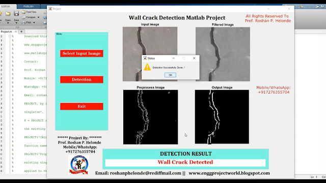
            Engineering structures like concrete surface, beams are often subjected to fatigue stress, cyclic loading, that leads to the cracks that usually initiate at the microscopic level on the structure’s surface. The cracks on the structure reduce local stiffness and cause material discontinuities. Early detection allows preventive measures to be taken to prevent damage and possible failure. Crack detection is the process of detecting the crack on the wall surface using image processing techniques. For fast and reliable surface defect analysis, Automatic crack detection is developed instead of the slower subjective traditional human inspection procedures. Thereby a safer survey methodology is adapted. Automatic crack detection is very effective for Non-destructive testing. Cracks on the concrete surface are one of the earliest indications of degradation of the structure which is critical for the maintenance as well the continuous exposure will lead to the severe damage to the environment. Manual inspection is the acclaimed method for the crack inspection. In the manual inspection, the sketch of the crack is prepared manually, and the conditions of the irregularities are noted. Since the manual approach completely depends on the specialist’s knowledge and experience, it lacks objectivity in the quantitative analysis. So, automatic image-based crack detection is proposed in this project. This project is developed in matlab.

PROJECT OUTPUT


PROJECT DEMO VIDEO

Contact:
Prof. Roshan P. Helonde
Mobile: +917276355704
WhatsApp: +917276355704
Email: roshanphelonde@rediffmail.com

Friday 16 February 2024

Medical Image Fusion Using DWT Discrete Wavelet Transform Matlab Project With Source Code | Final Year Project

ABSTRACT

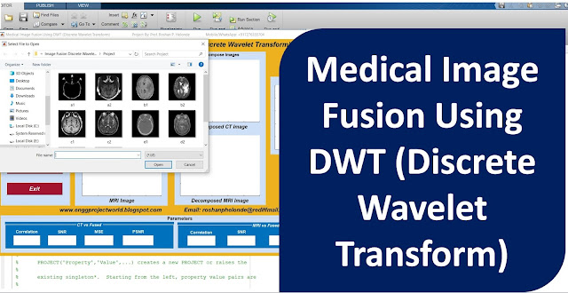
            Different medical imaging techniques such as X-rays, computed tomography (CT), magnetic resonance imaging (MRI) provide different perspectives for the human body that are important in the physical disorders or diagnosis of diseases .To derive useful information from multimodality medical image data medical image fusion has been used. In the medical field different radiometric scanning techniques can be used to evaluate and examine the inner parts of the body. The idea is to improve the image content by fusing images like computer tomography (CT) and magnetic resonance imaging (MRI) images, so as to provide as much details as possible for the sake of diagnosis. The objective of image fusion is to merge information from two images of the same image. The resultant image after image fusion is more suitable for human and machine perception and further helpful for image-processing tasks such as segmentation, feature extraction and object recognition. This project mainly presents image fusion using wavelet method for multispectral data and high-resolution data conveniently, quickly and accurately in MATLAB. Wavelet toolbox with abundant functions, provide a quick and convenient platform to improve image visibility. The work covers the selection of wavelet function, the use of wavelet based fusion algorithms on CT and MRI medical images, implementation of fusion rules and the fusion image quality evaluation. Matlab Results show that effectiveness of Medical Image Fusion with Wavelet Transform on preserving the feature information for the test images.

PROJECT OUTPUT


PROJECT DEMO VIDEO

Contact:
Prof. Roshan P. Helonde
Mobile: +91-7276355704
WhatsApp: +917276355704
Email: roshanphelonde@rediffmail.com

Wednesday 14 February 2024

Plant Disease Detection Using CNN Convolutional Neural Network Matlab Project With Source Code Final Year Project

 ABSTRACT

            Today's better technologies have enabled people to provide the adequate nutrition and food needed to meet the needs of the world's growing population. If we talk about India unequivocally, 70% of the Indian people is directly or by suggestion related to the cultivating territory, which remains the greatest region in the country. If we explore the broader Picture According to Research Conducted by 2050 overall yield creation can augment by at any rate half putting more weight on the inside and out pushed and cultivating Sector. The greater part of the Farmers is poor and have no inclination in development which may incite hardships more essential than half because of pets and sicknesses of plant. Vegetables and fruits are common items and the principal agricultural things. Powerful dependence on engineered pesticides achieves the high substance content which creates in the earth, air, water, and shockingly in our bodies antagonistically influence the environment. At present, the conventional technique of visual inspection in humans by visual inspection makes it impossible to characterize plant diseases. Advances in computer vision models offer fast, normalized, and accurate answers to these problems. Early Disease Detection and pets are important for better yield and quality of crops. With Reduction in Quality of the agricultural Product, Disease Plant can lead to the huge Economic Losses to the Individual farmers. In country like India whose major Population is involved in Agriculture It is very important to find the disease at early stages. Faster and precise prediction of plant disease could help reducing the losses. Image  processing  techniques involves in this project are  image  acquisition,  image  preprocessing,  image  segmentation and classification done using CNN Convolutional Neural Network. This project is developed in matlab software. Development of  automatic detection  system using advanced  computer technology  such as  image processing help to support  the  farmers in  the identification  of diseases at an early or initial stage and provide useful information for its control.

PROJECT OUTPUT


PROJECT DEMO VIDEO

Contact:
Prof. Roshan P. Helonde
Mobile: +91-7276355704
WhatsApp: +917276355704
Email: roshanphelonde@rediffmail.com