Tuesday 31 December 2019

Paddy Leaf Disease Detection using image processing matlab project with source code

ABSTRACT
              Agriculture is the main backbone for most of the developing/developed countries; agriculture production itself is the main feed for ever growing populations and it is the major source of income for the rural people/farmers especially in India. In India farmers are called “the backbone of India”. The main aim of the proposed system is to detect and classify the diseases in paddy leafs. Paddy Diseases Classification comprises of two steps: first one is Detection, Extraction and Segmentation of diseases. Secondly, Feature extraction, Classification and Grade the level of disease by using Support Vector Machine (SVM) classifiers respectively. The proposed system has been experimentally tested for our own dataset and results achieved are encouraging.
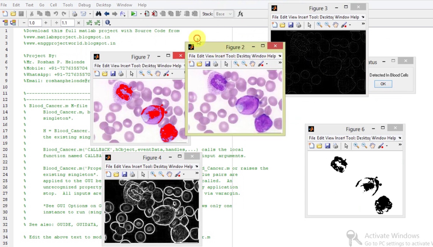
PROJECT OUTPUT

PROJECT VIDEO

Contact:
Mr. Roshan P. Helonde
Mobile: +91-7276355704
WhatsApp: +91-7276355704
Email: roshanphelonde@rediffmail.com

Wednesday 25 December 2019

Matlab code for Fake Currency Detection using Image Processing

ABSTRACT
                  The Reserve Bank is the one which issue bank notes in India. Reserve Bank, changes the design of bank notes from time to time. Reserve bank uses several techniques to detect fake currency. Common people faces many problems for the fake currency circulation and also difficult to detect fake currency, suppose that a common people went to a bank to deposit money in bank but only to see that some of the notes are fake, in this case he has to take the blame. As banks will not help that person. Some of the effects that fake currency has on society include a reduction in the value of real money; and inflation due to more fake currency getting circulated in the society or market which disturbs our economy and growth - an some illegal authorities an artificial increase in the money supply,a decrease in the acceptability of paper money and losses. Our aim is to help common man to recognize currency. Proposed system is based on image processing and makes the process automatic and robust. Shape information are used in our algorithm. Original Note Detection Systems are present in banks but are very costly. We are developing an image processing algorithm which will extract the currency features and compare it with features of original note image. This system is cheaper and can provide accuracy on the basics of visual contents of note.

PROJECT OUTPUT

PROJECT VIDEO

Contact:
Mr. Roshan P. Helonde
Mobile: +91-7276355704
WhatsApp: +91-7276355704
Email: roshanphelonde@rediffmail.com

Monday 23 December 2019

Finger Nail Disease Detection using Image Processing Matlab Project with Source Code

ABSTRACT
          This project gives idea to predict diseases using the colour of the nail at early stage of diagnosis. The main aim of our project is to analyze the disease without causing harm to humans. In earlier traditional system of disease detection, doctors observe the nails of patients and will predict the disease. Many diseases can be identified by analyzing nails of patients .But it is difficult for human eyes to differentiate the slight changes in colour. So it is less accurate and time consuming. Our proposed system can be quite useful to overcome this issue since it is fully computer based. The input to the proposed system is image of nail. The system will process the nail image and will extract the nail’s features to diagnose the disease. Human nail consist of various features, our proposed system uses nail color changes to diagnose the disease. Here, first training set data is prepared from nail images of patients with specific diseases. This training data set is compared with extracted feature from input nail image to obtain the result. In our experiment, we found that training set data are correctly matched with color feature of nail image results. It is focused on the system of image recognition on the basis of color analysis. The proposed system is based on the algorithm which automatically extracts only nails area from scanned back side of palm (Region of Interest). These selected pixels are processed for further analysis using median filters. The system is fully computer based, so even small discontinuities in color values are observed, and we can detect color changes in the initial stage of disease.

PROJECT OUTPUT

PROJECT VIDEO

Contact:
Mr. Roshan P. Helonde
Mobile: +91-7276355704
WhatsApp: +91-7276355704
Email: roshanphelonde@rediffmail.com

Tuesday 17 December 2019

Rain Removal using Image Processing Matlab Project with Source Code || IEEE Based Project

ABSTRACT
                 The rain removal from an image in the rainy season is also a required task to identify the object in it. It is a challenging problem and has been recently investigate extensively. In this project the entropy maximization and background estimation based method is used for the rain removal. This method is based on single-image rain removal framework. The raindrops are greatly differing from the background, as the intensity of rain drops is higher the background. The entropy maximization is very much suitable for the rain removal. Experimental results express the efficacy of the rain removal by proposed algorithm is better than the method based on saturation and visibility features. The rain and non-rain parts in a single image are very closely mixed up and the identification of rain streaks is not an easy task. In this project, we compare a single-image rain streak removal based on morphological component analysis (MCA) by decomposition of rain streaks. The signal and image processing for the filtering and region specification are discussed in the previous works. In this method, a bilateral filter is applied for an image to decompose it into the low-frequency (LF) and high-frequency (HF) parts. The HF part is then decomposed into rain component and non-rain component by performing sparse coding and dictionary learning on MCA.

PROJECT OUTPUT

PROJECT VIDEO

Contact:
Mr. Roshan P. Helonde
Mobile: +91-7276355704
WhatsApp: +91-7276355704
Email: roshanphelonde@rediffmail.com

Blood Cancer (Leukemia Cancer) Detection Using Image Processing Matlab Project with Source Code

ABSTRACT
        Blood cancer is the most prevalent and it is very much dangerous among all type of cancers. Early detection of blood cancer has the potential to reduce mortality and morbidity. There are many diagnostic technologies and tests to diagnose blood cancer. However many of these tests are extremely complex and subjective and depend heavily on the experience of the technician. To obviate these problems, image processing techniques is use in this study as promising modalities for detection of different types of blood cancer. The accuracy rate of the diagnosis of blood cancer by using the image processing will be yield a slightly higher rate of accuracy then other traditional methods and will reduce the effort and time. We first discuss the preliminary of cell biology required to proceed to implement our proposed method. This project presents a new automated approach for blood Cancer detection and analysis from a given photograph of patient’s cancer affected blood sample. The proposed method is using image improvement, image segmentation for segmenting the different cells of blood, edge detection for detecting the boundary, size, and shape of the cells and finally Clustering then final decision of blood cancer based on the number of different cells.

PROJECT OUTPUT

PROJECT VIDEO

Contact:
Mr. Roshan P. Helonde
Mobile: +91-7276355704
WhatsApp: +91-7276355704
Email: roshanphelonde@rediffmail.com

Saturday 14 December 2019

Blood Group Determination Using Image Processing Matlab Project with Source Code

ABSTRACT
          Determining of blood types is very important during emergency situation before administering a blood transfusion. Presently, these tests are performed manually by technicians, which can lead to human errors. Determination of the blood types in a short period of time and without human errors is very much essential. A method is developed based on processing of images acquired during the slide test. The image processing techniques such as Pre-processing, Segmentation, Thresholding, Morphological operations and Support Vector Machine are used. The images of the slide test are obtained from the pathological laboratory are processed and the occurrence of agglutination are evaluated. Thus the developed automated method determines the blood type using image processing techniques. The developed method is useful in emergency situation to determine the blood group without human error. The slide test consists of the mixture of one drop of blood and one drop of reagent, being the result interpreted according to the occurrence or not of agglutination. The combination of the occurrence and non occurrence of the agglutination determines the blood type of the patient. 

PROJECT OUTPUT

PROJECT VIDEO

Contact:
Mr. Roshan P. Helonde
Mobile: +91-7276355704
WhatsApp: +91-7276355704
Email: roshanphelonde@rediffmail.com

Saturday 7 December 2019

Plant Disease Detection & Classification using Neural Network Matlab Project with Source Code

ABSTRACT
            The detection and classification of plant diseases are the crucial factors in plant production and the reduction of losses in crop yield. This paper proposes an approach for leaf disease detection and classification on plants using image processing. The algorithm presented has three basic steps: Image Pre-processing and analysis, Feature Extraction and Recognition of plant disease. The plant disease diagnosis is restricted by person’s visual capabilities as it is microscopic in nature. Due to optical nature of plant monitoring task, computer visualization methods are adopted in plant disease recognition. The aim is to detect the symptoms of the disease occurred in leaves in an accurate way. Once the captured image is pre-processed, the various properties of the plant leaf such as intensity, color and size are extracted and sent to SVM classifier with Neural Network for classification. 

PROJECT OUTPUT

PROJECT VIDEO

Contact:
Mr. Roshan P. Helonde
Mobile: +91-7276355704
WhatsApp: +91-7276355704
Email: roshanphelonde@rediffmail.com

Friday 6 December 2019

Audio Steganography for Data Hiding Matlab Project with Source Code

ABSTRACT
          Information security is one of the most important factors to be considered when secret information has to be communicated between two parties. Cryptography and steganography are the two techniques used for this purpose. Cryptography scrambles the information, but it reveals the existence of the information. Steganography hides the actual existence of the information so that anyone else other than the sender and the recipient cannot recognize the transmission. In steganography the secret information to be communicated is hidden in some other carrier in such a way that the secret information is invisible. In this paper an image steganography technique is proposed to hide audio signal in image in the transform domain using wavelet transform. The audio signal in any format wav is encrypted and carried by the image without revealing the existence to anybody. When the secret information is hidden in the carrier the result is the stego signal. In this work, the results show good quality stego signal and the stego signal is analyzed for different attacks. It is found that the technique is robust and it can withstand the attacks. The quality of the stego image is measured by Peak Signal to Noise Ratio (PSNR), Mean Square Error. The quality of extracted secret audio signal is measured by Signal to Noise Ratio (SNR), Squared Pearson Correlation Coefficient (SPCC). The results show good values for these metrics.

PROJECT OUTPUT

PROJECT VIDEO

Contact:
Mr. Roshan P. Helonde
Mobile: +91-7276355704
WhatsApp: +91-7276355704
Email: roshanphelonde@rediffmail.com

Brain Tumor Detection using Rough Set Theory Algorithm Matlab Project with Source Code

ABSTRACT
              Brain tumor is a life threatening disease and its early detection is very important to save life. The tumor region can be detected by segmentation of brain Magnetic Resonance Image (MRI). Once a brain tumor is clinically suspected, radiologic evaluation is required to determine the location, the extent of the tumor, and its relationship to the surrounding structures. This information is very important and critical in deciding between the different forms of therapy such as surgery, radiation, and chemotherapy. The segmentation must be fast and accurate for the diagnosis purpose. Manual segmentation of brain tumors from magnetic resonance images is a tedious and time-consuming task. Also the accuracy depends upon the experience of expert. Hence, the computer aided automatic segmentation has become important. MRI scanned images offer valuable information regarding brain tissues. MRI scans provide very detailed diagnostic pictures of most of the important organs and tissues in our body. It is generally painless and noninvasive. It does not produce ionizing radiation. So MRI is one of the best clinical imaging modalities. Several automated segmentation algorithms have been proposed. But still segmentation of MRI brain image remains as a challenging problem due to its complexity and there is no standard algorithm that can produce satisfactory results. The aim of this research work is to propose and implement an efficient system for tumor detection and classification. The different steps involved in this work are image pre-processing for noise removal, feature extraction, segmentation and classification.

PROJECT OUTPUT

PROJECT VIDEO

Contact:
Mr. Roshan P. Helonde
Mobile: +91-7276355704
WhatsApp: +91-7276355704
Email: roshanphelonde@rediffmail.com

Thursday 5 December 2019

Bone Fracture Detection using Image Processing Matlab Project with source Code

ABSTRACT
         Analysis of medical images plays a very important role in clinical decision making. For a long time it has required extensive involvement of a human expert. However, recent progress in data mining techniques, especially in machine learning, allows for creating decision models and support systems that help to automatise this task and provide clinicians with patient-specific therapeutic and diagnostic suggestions. In this project, we describe a study aimed at building a decision model (a classifier) that would predict the type of treatment (surgical vs. non-surgical) for patients with bone fractures based on their X-ray images. We consider two types of features extracted from images (structural and textural) and used them to construct multiple classifiers that are later evaluated in a computational experiment. Structural features are computed by applying the Hough transform, while textural information is obtained from Gray-level occurrence matrix. In research reported by other authors structural and textural features were typically considered separately. Our findings show that while structural features have better predictive capabilities, they can benefit from combining them with textural ones.

PROJECT OUTPUT

PROJECT VIDEO

Contact:
Mr. Roshan P. Helonde
Mobile: +91-7276355704
WhatsApp: +91-7276355704
Email: roshanphelonde@rediffmail.com

Matlab Code for Blood Group Detection Using Image Processing

ABSTRACT
          Determining of blood types is very important during emergency situation before administering a blood transfusion. Presently, these tests are performed manually by technicians, which can lead to human errors. Determination of the blood types in a short period of time and without human errors is very much essential. A method is developed based on processing of images acquired during the slide test. The image processing techniques such as thresholding and morphological operations are used. The images of the slide test are obtained from the pathological laboratory are processed and the occurrence of agglutination are evaluated. Thus the developed automated method determines the blood type using image processing techniques. The developed method is useful in emergency situation to determine the blood group without human error. The slide test consists of the mixture of one drop of blood and one drop of reagent, being the result interpreted according to the occurrence or not of agglutination. The combination of the occurrence and non occurrence of the agglutination determines the blood type of the patient. 

PROJECT OUTPUT

PROJECT VIDEO

Contact:
Mr. Roshan P. Helonde
Mobile: +91-7276355704
WhatsApp: +91-7276355704
Email: roshanphelonde@rediffmail.com

Tuesday 3 December 2019

Face Recognition Based Attendance Maintenance Matlab Project with Source Code

ABSTRACT
               In this project we are proposing an automatic attendance system which can be used in every organisation to mark the attendance of employees. The main application of Automatic attendance system is seen in teaching institutions, where the attendance of students has to be regularly monitored on daily basis. The method developed provides a secure and effective may recording attendance. Automatic face recognition (AFR) technologies have made many improvements in the changing world. Smart Attendance using Real-Time Face Recognition is a real-world solution which comes with day to day activities of handling student attendance system. Face recognition-based attendance system is a process of recognizing the students face for taking attendance by using face biometrics based on high - definition monitor video and other information technology. In my face recognition project, a computer system will be able to find and recognize human faces fast and precisely in images or videos that are being captured through a surveillance camera.

PROJECT OUTPUT

PROJECT VIDEO

Contact:
Mr. Roshan P. Helonde
Mobile: +91-7276355704
WhatsApp: +91-7276355704
Email: roshanphelonde@rediffmail.com

Monday 2 December 2019

Osteoporosis Detection using Image Processing full Matlab Project with source code

ABSTRACT
               Osteoporosis is a progressive bone disease that is characterized by a decrease in bone mass and density which can lead to an increased risk of fracture. Osteoporosis is a state of having brittle and fragile bone which arises due to vitamin deficiency, tissue loss, hormonal changes. Osteoporosis can be efficiently detected by calculating various features like Bone mineral density (BMD), statistical features from various trabecular region such as hip, toe, elbow, etc. Detection of bone disorders are done with the help of bone densitometer. The bone densitometer uses a technique that the bone density can be measured in-terms of Tscore. Osteoporosis is a condition in which the bone becomes porous and fragile due loss in bone mineral density and gets more susceptible to fracturing. osteopenia refers to early signs of bone loss that can turn to osteoporosis. Both osteoporosis and osteopenia are increasingly found in aging women who have attained their menopause. The symptoms of osteoporosis include pain in the bones, or lower back, bone fracture and
loss of height over a course of time.

PROJECT OUTPUT

PROJECT VIDEO

Contact:
Mr. Roshan P. Helonde
Mobile: +91-7276355704
WhatsApp: +91-7276355704
Email: roshanphelonde@rediffmail.com