ABSTRACT
Brain is the kernel part of the body. Brain has a very complex structure. Brain is hidden from direct view by the protective skull. This skull gives brain protection from injuries as well as it hinders the study of its function in both health and disease. But brain can be affected by a problem which cause change in its normal structure and its normal behavior .This problem is known as brain tumor. Brain tumor causes the abnormal growth of the cells in the brain. The cells which supplies the brain in the arteries are tightly bound together thereby routine laboratory test are inadequate to analyze the chemistry of brain. Brain tumor diagnosis is a very crucial task. This system provides an efficient and fast way for diagnosis of the brain tumor. Proposed system consists of multiple phases. First phase consists of texture extraction from brain MR images. Second phase classify brain images on the bases of these texture feature using deep learning classifier. After classification tumor region is extracted from those images which are classified as malignant or benign. Segmentation consists of tumor extraction phases. This project is developed in matlab using deep learning cnn.
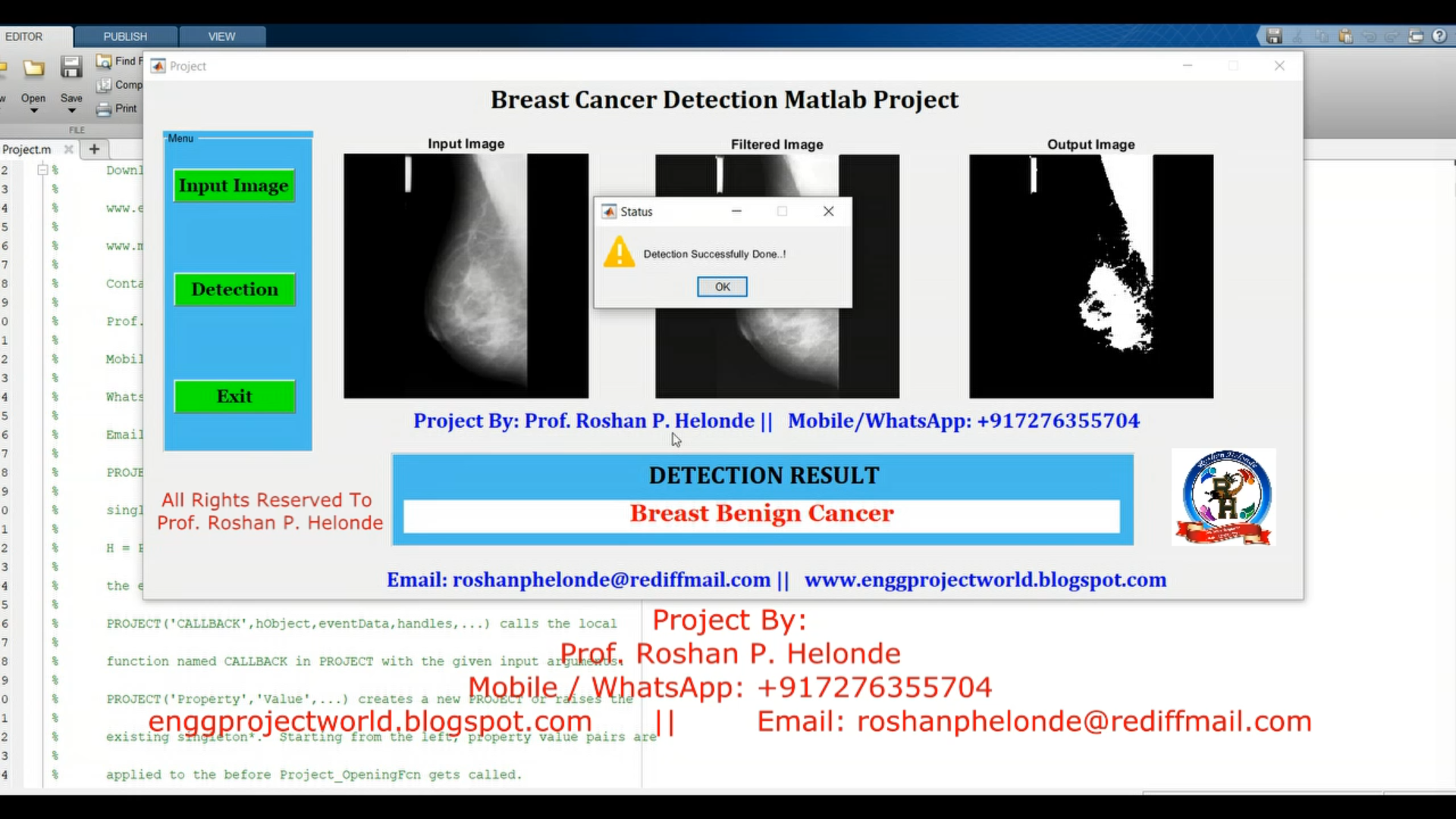
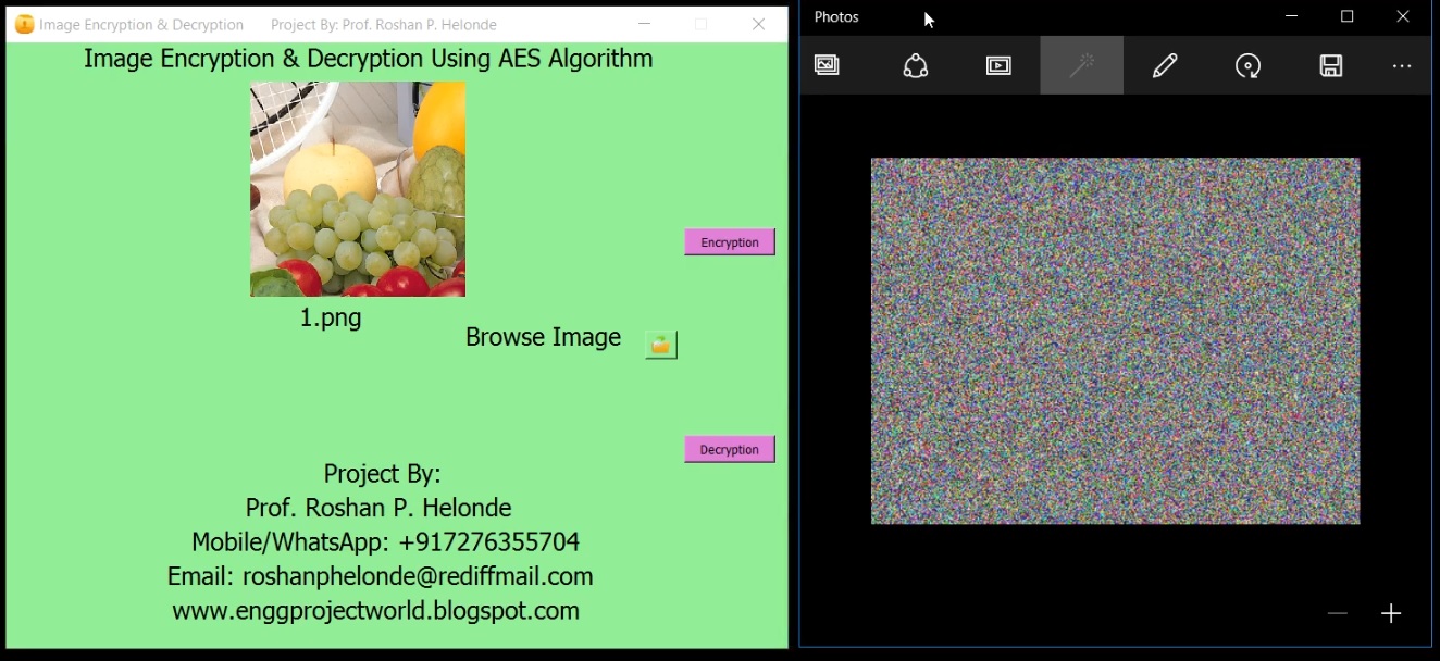
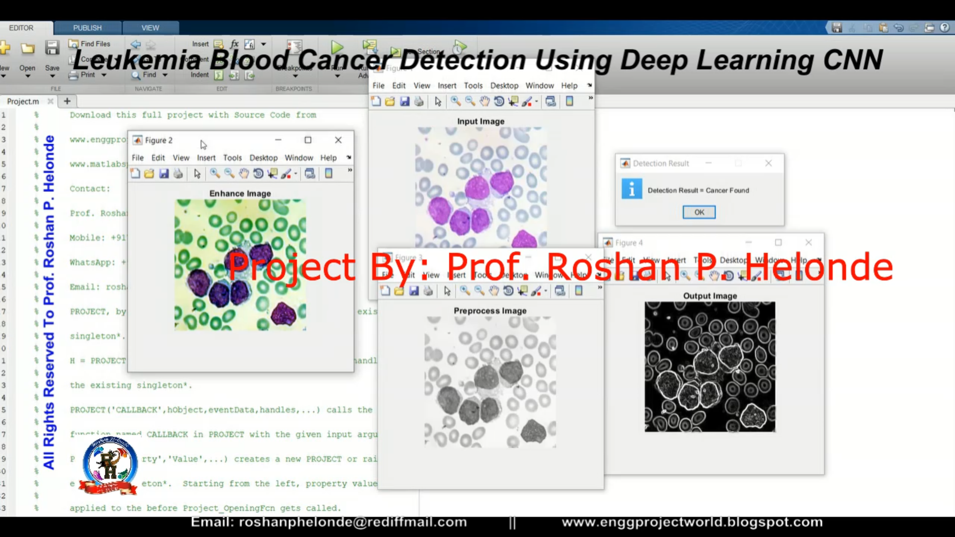
PROJECT OUTPUT
.png)
PROJECT DEMO VIDEO
.png)

.png)


.png)
.png)


.png)
.png)

.png)